What do you know about Molars? The human dentition has a clear structure. Normally, after its complete formation, there should be 32 teeth in the oral cavity: sixteen on the lower jaw and the same number on the upper. Moreover, all of them have their structure, depending on what functions they perform and where exactly they are located. For example, fangs and incisors are designed to bite, tear, and hold food. Premolars perform similar tasks, plus they are responsible for chewing operations. There is a fourth type – molar root units. Leading specialists of Dentika dentistry will tell you in detail about molars – what they are, what work teeth do, and how they look in adults and children.

Location and anatomical features
Man is an omnivore. That is, for the normal course of all metabolic processes in the body, he needs a variety of food, including coarse, vegetable, and animal origin. Bite and swallowing the product is not enough. For it to be assimilated, high-quality chewing is required. It is for this that the units of dentition are responsible, which are assigned the numbers 6, 7, and 8 in mature people and 4, and 5 in paediatric patients. They are called molars.
Their chewing area is 1 cm2. They can withstand the pressure of 70-75 kg. It turns out that after the complete formation of the jaw in the mouth, there are only 12 of them: 3 on top and bottom, on each side. The latest, the third in a row, are called wisdom teeth. They erupt in adults, but often they do not happen at all. The root part can have 2-3 or more branches.
Structural features
Those dental units that are located below, as a rule, have 3 canals and 2 roots, those that are at the top have 4 and 3, respectively. The extreme ones differ in a more significant surface area and slightly different anatomy.
The size of molar crowns in diameter reaches 7-10 mm. They are diamond-shaped with rounded edges. There are 4 tubercles on the surface, separated by three grooves located transversely.
There are several types of roots:
- Palatine;
- Bucco-distal;
- Bucco-mesial.
The latter is the largest. The first ones are medium in size. The second variety will be the shortest. Each subsequent tooth has a smaller root and crown part. Thus, the 1st is the largest, and the middle ones are smaller. Extreme has no predecessors. Often come into contact with 2 antagonists. There is nothing pathological in this. Such a scheme is considered quite normal.
Internal structure
Molars are made up of the following elements:
- The outer shell is enamel. It is considered the hardest structure in the human body. Acts as a protection against injury and penetration of pathogenic microorganisms.
- Dentine. There are channels on it through which nutrients enter.
- The nerve is the pulp. This is a neurovascular bundle that is responsible for nutrition and blood supply, and the formation of secondary dentin tissue. Located in the pulp chamber and tubules.
- Root. The upper ones have 3 + 4 grooves. The lower ones have 2 + 3 channels.
- The tissue around the root is the periodontium. They hold the crown inside and absorb the load during the chewing process.
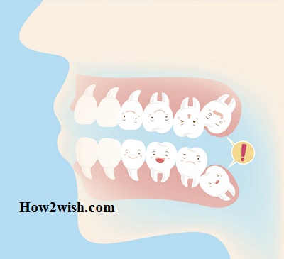
In the photo, you can see what kind of teeth the molars are and where they are in children.
How to Remove Tooth Abscess and Prevent It From Returning
Differences in structure depending on serial numbers
Depending on the order of eruption and location, dental units are usually classified into 1st, 2nd, and 3rd.
First
As mentioned earlier, they are the largest. They have the most extensive crown area (cubic, slightly elongated along the jaw row) and the length of the roots. Moreover, those that are on top have a more powerful root system than their antagonists from below.
Second
Several smaller sizes. The top can have any shape. The lower ones are always in the form of a cube. They have a clear cruciform groove that divides the surface into 4 tubercles.
Third root molars
Anatomical features:
- The crown part with a different area, and the roots of different lengths (but not too large, often they do not grow in depth, but to the side);
- Below they are larger than above;
- A large number of root branches (up to five), and they can grow together in pairs into one;
- On the surface, as a rule, there are three tubercles – two buccal and one lingual.
They appear only in adulthood. That is why they are called wisdom teeth.
When are the first primary molars formed?
They begin to erupt in childhood. Since the body of each child is individual, this can occur at different times – usually in the period from 1 year to 2.5 years. They hatch quite painfully.
By the time they appear, the chewing reflex in children is already developed. Without these dental units, solid food intake was problematic. Therefore, the kids immediately begin to use them, because without them they experienced some discomfort. Because hard food is introduced into the diet, the formation of the jaw is accelerated.
With a milk bite, there are eight molars in the oral cavity. Although they are behind the fangs, they appear earlier. They are cut in pairs, first the lower one, followed immediately by the upper one.
The second molar units hatch after two years. In some cases, this may happen sooner or later. This is considered normal. This may be due to hereditary predisposition.
Since these chewing organs have a vast surface and a large root part, their eruption is quite painful. This period, probably, remains in the memory of every parent. Only a small percentage of babies do not experience any discomfort. Piping is accompanied by the following symptoms:
- Swelling and redness of the gums;
- Increased salivation;
- Restless behaviour;
- Worsening sleep;
- Refusal of food;
- Increase in body temperature;
- Runny nose (if immunity is weakened in parallel);
- Diarrheal (in some cases).
Note! When a child begins to cut teeth, it must be shown to a paediatric dentist. This will allow you to detect problems promptly, if any, and begin to correct them.
Canines, incisors, and premolars also begin to actively grow at the age of about 24 months. By 2.5 years, as a rule, the entire set of milk bites appears.
Replacement with permanent chewing teeth occurs between the ages of 5 and 12, with the molars being the first to fall out. The drop order is opposite to the spawn pattern. You can understand that a dental unit will soon fall out by loosening it. It is not recommended to delete it by force. She must fall on her own. Otherwise, the replacement tooth may grow out of place, in the wrong place.
Signs of permanent occlusion formation
It is possible to determine that the root chewing organs will soon appear even before the milk ones fall out. The following symptoms are observed:
- Expansion of the jaw, which is accompanied by the appearance of gaps in the row;
- The appearance of sufficient space behind the extreme lateral milkers;
- Swollen gums.
Rarely, there is an increase in temperature or a deterioration in well-being. In most cases, parents do not even notice that a new one has appeared in place of the missing tooth unit.
Is it possible to loosen temporary molars?
The process of shedding begins with the softening of the root. This happens as a result of the growth of the jaw: the free space for the root chewing organs increases. They begin to take their place even at the moment when the dairy “sits” in their holes.
Therefore, the specialists of the dental clinic “Dentika” do not recommend loosening the predecessors. If they fall prematurely, jawbone development may be halted. Because of this, all permanent dental units simply will not fit. They will begin to shift, grow crookedly, breaking out of the general row.
Helping a child change teeth
When crowns with sharp edges cut the gum to get out, naturally, this is accompanied by painful sensations. If the growth of fangs begins in parallel, then this can cause even more significant discomfort.
To alleviate the condition, the doctor may prescribe painkillers designed specifically for children that will not harm the children’s bodies. Also in the pharmacy, you can buy specialized rubber rings with liquid inside (teethers). They allow you to relieve pain.
Since wounds appear during the change of bite, the risk of infection increases. Therefore, it is recommended to rinse your mouth several times a day, for example, with soda solution or chamomile. You should not expect any complications if you regularly (at least 2 times a year) show your child to the dentist, and follow the hygiene of the oral cavity.
Important! Any drugs and folk remedies should be prescribed exclusively by a doctor. Their use without prior consultation with a specialist may be unsafe for the body.
When Dairy Becomes Indigenous
Replacement of temporary molars with permanent ones occurs after 6-7 years. As mentioned earlier, they form in the gum long before the milk jug falls out. Even if the child has reached the age when a change should be observed, but it is not there, this process cannot be accelerated. Otherwise, then the jaw may remain underdeveloped, and the dental units will grow crookedly on the second row.
Cutting pattern:
- Sixes – 6-8 years;
- Upper and lower sevens – 12-13 years;
- Large molars (these are wisdom teeth) – from 17 to 30 years or more.
The latter may not appear at all and remain inside the gums. Sometimes they may need to be surgically removed. The decision is made by the doctor. When the bite changes, it is necessary to visit the dental office regularly. This will help to detect problems promptly and fix them.
Indigenous dental units and prevention of their loss
The permanent masticatory organs are stronger than the temporary ones. However, care should never be neglected. If they fall out, then new ones will not grow in their place. Preventive measures are as follows:
- Daily cleaning of the oral cavity 2 times a day (morning and evening);
- Using a thread or a special rinse after each meal;
- Regular visits to the dentist, even if there are no complaints (at least once every six months);
- Compliance with all recommendations of a specialist;
- Timely treatment of existing diseases;
- Avoidance of injury.
If there are already problems, the doctor will prescribe an effective treatment regimen. Conservative and surgical methods can be used. If the dental unit still could not be saved, you will have to resort to prosthetics. Modern dentistry is very advanced. Thanks to innovative technologies, implants, and crowns are visually indistinguishable from real masticatory organs. After their engraftment, the patient does not experience any discomfort.
The most common lesion is caries. This disease is characteristic of absolutely all teeth, but the molars are affected by its special form – fissure. The presence of grooves and depressions on the surface leads to the fact that food debris and bacteria accumulate inside and have a destructive effect on the enamel.
As a preventive measure, fissure sealing is performed. During the procedure, the grooves and depressions are sealed with a specialized sealant. The therapy is carried out according to the standard scheme – the affected areas are cleaned, and a filling is performed.
If you do not start treating caries promptly, it will lead to complications in the form of:
- Pulpitis (inflammatory process in the pulp);
- Periodontitis (inflammation in the root zone).
In advanced cases, endodontic treatment may be required, which includes the following activities:
- Removal of pulp tissue;
- Forced expansion of channels and their disinfection;
- Filling with gutta-percha.
If conservative therapy does not bring the desired result and the lesion extends to the root, surgical intervention is prescribed (resection, hemisection, separation, amputation).
Periodontitis rarely affects molars, but dentists can diagnose it. This lesion is characterized by an inflammatory process in the ligaments and tissues that surround the root part. For a cure, complex therapeutic measures are prescribed, including professional cleaning and hygiene procedures, curettage, and antibacterial and anti-inflammatory therapy.
The task of the dentist in this case is to preserve the molar units. They are responsible for the chewing process. Without them, the load will be unevenly distributed, which is why the development of dementia is possible.
Differences between molars and other teeth
Each of them has its purpose, respectively, different types have features. In molar varieties, they are as follows:
- The largest size of the crown and root part in the row;
- The upper ones have more channels than the lower ones;
- Those located below have 2 roots, and those located at the top have 3-4;
- The most extreme appear in adulthood, up to about 30 years, sometimes even later, but may not erupt at all;
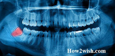
- Figure eights often do not come out completely (retention), and grow outside the jaw arch (dystopia).
Note! In 8-ok, the roots can be curved, often they are intertwined with each other. Therefore, they are the most difficult to treat, often easier to remove than cure.
Summing up
We told what molar teeth are, what they are in a row when they appear, and what it is accompanied by. They have their characteristics, which are due to their purpose. Undoubtedly, dental units are extremely important in the oral cavity, but molars are especially important. They are responsible for thoroughly chewing food. Without them, this task will fall on the neighboring chewing organs, and they will not be able to cope with such a load. This will lead to their destruction and premature loss. Therefore, if you suspect various violations, you should immediately contact your dentist. Make an appointment at our dental clinic “Dentika”!